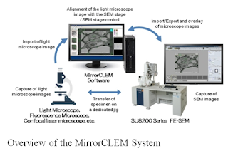
New MirrorCLEM System for Correlative Light and Electron Microscopy
At the center of stakes, we have the advancement of CLEM analysis in a variety of fields including medical and life sciences. We have: simplified correlative imaging of one location with both light and scanning electron microscopes.
 Based on the reality that, the observation of the same field in a specimen
with microscopes at different magnifications and observation characteristics
remained a difficult task; the MirrorCLEM system that Hitachi High-Tech and
RIKEN subsequently developed is an interesting curve to address the issue by supporting
quick and accurate CLEM analysis by using a FE-SEM.
For those who are unfamiliar, various
types of microscopes are used in a wide variety of fields such as
nanotechnology, materials, medical and life sciences. In the medical and life
sciences field in particular, SEMs are used to clarify the ultrastructure of
cells and tissues, while a type of light microscope called fluorescence microscopes
are being used increasingly to observe the localization and behavior of
proteins at the molecular level.

